The bite analysis that dentists have to look at
2022-08-09
Occlusal contact interference can occur in natural teeth or restorations, that is, it prevents contact between occlusal teeth that induce flexible movement when the occlusal or eccentric position is in motion.
Occlusal interference during the masticatory cycle interferes with the integration of muscle contractions, misaligns or distorts the jaw, and shifts the temporomandibular joint out of position.
In addition, the torque produced by the reduced efficiency of the lever mechanism can cause excessive force to be applied to the ligaments and adjacent muscle groups (especially the wing muscles).
The patterns of occlusal contact in occlusal interference include the following categories.
(1) centric relation premature contact (centric relation premature contact)
Even when the oral-maxillary system is functioning normally and the bite is normal, the lower jaw will gradually glide along one or more teeth during mid-early contact in the median position, such as maximizing the bite toward the median position.
The result of normal oblique motion is usually the mesial bevel of the lingual tip of the maxillary premolars or the distal bevel of the lingual tip of the mandibular premolars or molars.
There is much debate as to whether this early contact is pathological or physiological, but according to clinical studies, the more the medial occlusion between the teeth becomes more laterally larger 1mm in front of the normal medial contact, the more the temporomandibular joint becomes pathologically elongated.
Two types of occlusal interference can be observed.
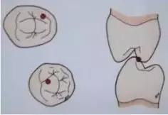
Figure 1. Occlusal interference in central position 1 - Interference between the support bevel of the maxillary support tooth tips.
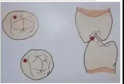
Figure 2. Occlusal interference in central position 2 - Interference between the induced surface of the mandibular supporting tip and the induced bevel of the maxillary leading tip.
(2) Centric occlusion early contact
Healthy natural dentition usually occurs 1mm in front of the median early contact, and the resting jaw muscles engage in the movement, closing the jaw at the median bite 0.5mm in front of the central condyle.
If the maximum occlusion begins laterally or in front of the median occlusion with mild early contact, due to the adaptability of the closed mouth muscles, the normal resting period of tension will be maintained at this time, but the non-tensive occlusal path will be followed.
However, in the case that the lateral anterior early contact is too large to make the median bite deviate, the mandibular slow closure will slide towards the median bite due to strong early contact from the rest period. However, in order to avoid such impact and slide, the muscle group will induce the avoidant closure path around the early contact.
Therefore, muscles adapt to the appropriately expanded avoidance pathway during mastication. In order to maintain the asymmetric median occlusal position at the position of maximum occlusal force, muscles will remain tense for a long time, eventually leading to condyle deviation to pathological median occlusal position, muscle overtension, muscle fatigue, muscle spasm and other diseases.
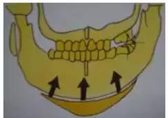
Figure 3. Closure from rest position to early contact
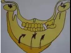
Figure 4. The mass of the temporomandibular joint induced by the deflection of the mouth from early contact to the maximum occlusal site.
(3) interference during eccentric movement
A. Working side interference
Interference caused by tooth induced eccentric mandibular motion, between the induced surface of the mandibular molar supporting tooth tips and the induced bevel of the maxillary molar tips or in the buccal bevel of the mandibular lingual tips, may interfere with dental function or fang-induced on the working side.
This manipulation interferes with the working side and condyle-induced dissonance of the teeth during lateral movement, thus damaging the joint surface and causing excessive force to extend the ligaments.
In addition, different levers and directions of forces are induced from non-working side disturbances, which can lead to chaotic neuromuscular phenomena. Working side interference with strong muscle contractions can also cause jaw distortion and deformation.
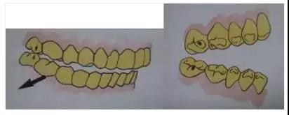
Figure 5. Working side interference between buccal bevel of mesial buccal cusp of mandibular second molar and lingual bevel of mesial buccal cusp of maxillary second molar.
B. Non-working side interference
The possible non-working side interference during lateral movement occurs between the buccal bevel of lingual apex of maxillary large and minor molars and the lingual bevel of mandibular buccal apex, which will interfere with the mechanism of contact induction on the working side. Such non-working side (equalizing side) interference will bring more damage to temporomandibular joint than the working side interference.
Non-working side contact may interfere with the coordination of the non-working condyles in order to maintain the spacing between the non-working teeth, may cause dislocation of the condyles with extension of the non-working ligament, or may distort the condyles and cause trauma to the joint.
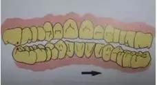
Figure 7. Non-working lateral interference between the lingual bevel of the distal buccal cusp of the mandibular second molar and the buccal bevel of the mesial lingual cusp of the maxillary second molar.
C. Interference with forward motion
Anterior motion depends on the coordination between the tooth induction function and the anterior condyle induction to maintain the normal induction effect. The interference that may occur during the extension movement can be divided into the anterior interference similar to the working interference in the anterior teeth and the non-working interference in the rear molars.
* front interference
Due to the interference between the cross section of the mandibular anterior teeth and the induction slope of the maxillary anterior teeth, the exact forward induction is hindered during the extension movement, which is generally caused by the poor appearance of the lingual surface of the prosthesis of the maxillary anterior teeth, the inappropriate position of the mandibular cross section, and the inappropriate maintenance contact in the anterior teeth.
Due to this occlusal phenomenon, the extension is induced by the contact of one or two teeth. Without proper anterior induction and coordination, the remaining teeth cannot be separated.
This contact is called forward induced interference.

Figure 8. Anterior interference in the anterior teeth
* Rear interference
The induction of the extension motion induced by contact between the distal bevel in the maxillary molars and the mesial induction in the mandibular molars supporting the tooth tips should be maintained precisely. Interference that interferes with the direction of this induced molars is called posterior interference. This is often the case when the occlusal surface of a lost molar or adjacent molar overerupts or shifts in position, and the occlusal plane of a damaged molar is not changed.

Figure 9. Difficulty opening the mouth due to anterior induced interference in the upper and lower second molars.
The mesial buccal bevel of the distal buccal cusp of the mandibular molars is in contact with the distal palatal bevel of the mesial buccal cusp of the maxillary molars.
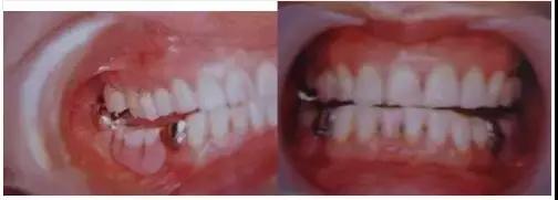
FIG. 10. Anterior induced interference caused by posterior molar interference after the completion of a repair system that did not consider the destruction of the occlusal plane of the mandibular third molar.
The bite is highly uncoordinated
A. Height reduction
As the posterior molar occlusion triggered by the loss of normal posterior molar occlusal support, partial occurrence of the molar is called posterior bite collapse.
Loss of molar occlusal support is gradually initiated by tooth movement of the remaining teeth and improper dental prosthesis. In the case of complete loss of the left and right molars and premolars, the mandibular condyles are dislocated in the middle and upper reaches of the mandibular condyles when the mouth is closed in the direction of the contact between the teeth in front of the mandibular teeth that tilt at the lingual surface of the maxillary anterior teeth.
In patients with complete dentures, a lower setting of the occlusal height can cause contraction of the occlusal muscle, sometimes causing fascia pain.
B. The height increases
This is a condition in which the occlusal height increases so much that freeway space can be closed off as a result of the constructed prosthesis, which can cause excessive muscle elongation, muscle overtension, muscle fatigue, and pain.

Figure 11. Pathological conditions of temporomandibular joint site and muscular system induced by molar occlusal collapse。

Figure 12. Excessive free separation can also trigger design-related muscle contractions


